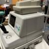
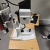
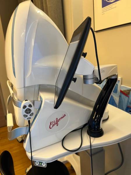
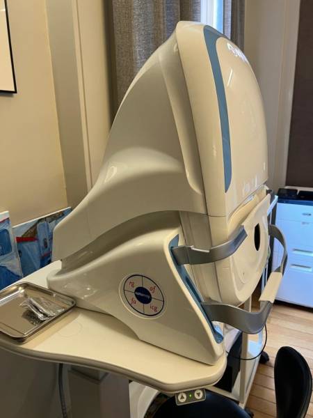
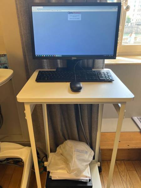
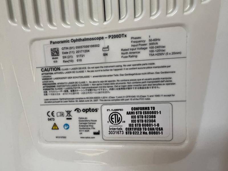


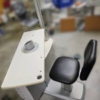
2017 OPTOS California
$55,000.00
It is the high end model of the product range, icg, it offers the following features (Thanks to the manufacturer brochure – available on demand)
The only single-capture ultra-widefield retinal image as defined by The International Widefield Imaging Study Group.
Non-mydriatic retinal imaging in less than ½ second has been shown to decrease patient visit time, enable doctors to see 7% more patients, and help doctors visualize pathology outside of the view of traditional small field fundus photography.
cSLO technology images through most cataracts5 and small pupils (2 mm).
Color rg mode produces 3 images in a single capture: color rg, sensory red-free, and choroidal.
Green laser af shows details across the entire retina.
Image overlay tool facilitates comparison of images in different image modes and from visit to visit.
OptosAdvance™ Image Management software streamlines image review, referrals, and consultations.
DICOM compatible software supports compliance with the Code of Federal Regulations.
Accurate distance (mm) and area (mm2) measurements provide objective assessment of change over time.
Stereo disc imaging allows accurate assessment of the optic nerve to diagnose and follow the progression of glaucoma.
Auto-montage combines an optomap into a single image showing up to 220° (97%) of the retina
Imaging Modalities :
Color rg
Sensory (red free)
Choiroidal
Autofluorescence
Fluorescein Angiography
Icg Angiography
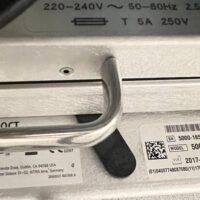
The device was under fuill maintenance control
Be the first to review “2017 OPTOS California”
Related products


Ophthalmology
$28,000.00


Ophthalmology
$3,200.00


Ophthalmology
$21,000.00


Ophthalmology
$10,000.00


Ophthalmology
$2,700.00

Ophthalmology
$6,000.00


Ophthalmology
$21,000.00


Ophthalmology
$90,000.00


Ophthalmology
$17,000.00


$9,500.00

Ophthalmology
$21,000.00


Ophthalmology
$1,500.00


Ophthalmology
$32,000.00


Ophthalmology
$6,500.00


Ophthalmology
$52,000.00


Ophthalmology
$63,000.00

Ophthalmology
$2,500.00


Ophthalmology


Ophthalmology
$11,000.00

Ophthalmology
$33,000.00


Ophthalmology
$8,000.00


Ophthalmology
$26,000.00


Ophthalmology
$3,800.00


Ophthalmology
$12,000.00
Reviews
There are no reviews yet.